
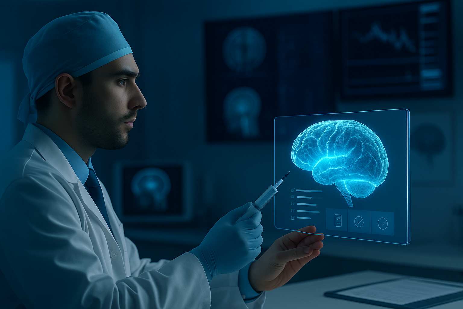
Advanced brain imaging plays a critical role in modern neurosurgery by providing detailed and precise visualization of brain structures. These imaging techniques enhance the surgeon’s ability to accurately diagnose and plan complex procedures, reducing risks and improving patient outcomes. By integrating technologies like functional MRI, diffusion tensor imaging, and AI-driven analysis, neurosurgeons achieve higher precision in targeting critical brain areas while preserving vital functions.
The continued development of imaging tools now allows for better tumor characterization, functional brain mapping, and real-time intraoperative visualization. This precision is essential in minimizing damage to healthy tissue and in handling complex cases such as brain tumors, epilepsy, and vascular abnormalities. With these advancements, neurosurgery is becoming progressively safer and more effective, driven by the improved clarity these imaging technologies provide.
These innovations also extend into augmented reality and multimodal imaging integration, which offer surgeons enhanced navigation during procedures. Such tools support decision-making by combining different data sources, further refining the surgical approach. The result is a new standard in brain surgery where accuracy and safety are significantly elevated by advanced imaging capabilities.
Overview of Advanced Brain Imaging in Neurosurgery
Advanced brain imaging in neurosurgery involves precise visualization techniques that enhance diagnosis, surgical planning, and intraoperative guidance. These methods improve the understanding of brain anatomy and function, supporting safer and more effective interventions.
Defining Advanced Brain Imaging
Advanced brain imaging refers to techniques that provide detailed structural and functional views of the brain. These include MRI, functional MRI (fMRI), diffusion tensor imaging (DTI), intraoperative MRI (iMRI), and CT scans.
Each technology offers specific insights. For example, fMRI identifies active brain regions, aiding functional mapping. DTI traces white matter pathways, critical for avoiding key neural tracts during surgery. iMRI allows real-time imaging during procedures, increasing precision.
Together, these modalities enable surgeons to accurately locate lesions, understand brain function, and monitor progress during surgery, reducing risks and improving outcomes.
Evolution of Imaging Techniques
Brain imaging has evolved significantly since the introduction of the radiograph in 1895. Early methods offered only rudimentary views, limiting neurosurgical precision.
The development of MRI revolutionized brain imaging by providing high-resolution, non-invasive images of soft tissues. Functional MRI added the ability to map brain activity. DTI further advanced imaging by visualizing nerve fiber pathways.
More recently, intraoperative imaging and augmented reality assist surgeons during operations, integrating live imaging with preoperative data. The use of artificial intelligence also begins to optimize image analysis, enhancing accuracy and surgical planning.
Challenges in Imaging for Neurosurgery
Despite advances, challenges remain in brain imaging for neurosurgery. One challenge is differentiating tumor tissue from healthy brain matter, especially in gliomas with blurred margins.
Functional imaging can be limited by patient movement or individual variability in brain organization. Real-time imaging requires balancing resolution with the need for quick updates during surgery.
Another challenge is integrating multi-modality images into coherent, actionable data for surgeons. Cost and accessibility of advanced imaging technologies also affect widespread adoption in clinical settings.
Continued innovation aims to address these issues, improving the reliability and utility of imaging in neurosurgical care.
| Challenge | Impact | Current Solutions |
| Tissue differentiation | Risk of incomplete resection | Advanced contrast agents, AI |
| Patient variability | Accurate functional mapping | Personalized imaging protocols |
| Real-time imaging speed | Intraoperative decision delays | Faster acquisition techniques |
| Data integration | Complexity in planning | Multimodal software platforms |
| Cost and access | Limited availability | Research into cost-effective tech |
Key Technologies in Advanced Brain Imaging
Advanced brain imaging in neurosurgery relies on a set of precise modalities that provide structural and functional details critical for surgical planning and intraoperative guidance. These technologies allow surgeons to identify anatomical landmarks, assess neural pathways, and evaluate brain activity related to essential functions.
Magnetic Resonance Imaging (MRI)
MRI produces high-resolution images by using magnetic fields and radio waves to visualize brain anatomy without ionizing radiation. It excels at detecting soft tissue contrast, enabling clear differentiation between gray and white matter, tumors, edema, and vascular structures.
In neurosurgery, MRI is essential for preoperative planning. It delivers detailed brain maps that help identify lesions and assess their proximity to critical areas. It also assists in monitoring postoperative changes and detecting complications like hemorrhage or ischemia.
MRI’s versatility extends to various sequences—such as T1, T2, and FLAIR—that highlight different tissue properties. These sequences guide surgeons in delineating tumor borders and planning minimally invasive approaches.
Diffusion Tensor Imaging
Diffusion Tensor Imaging (DTI) measures the diffusion of water molecules along white matter tracts, revealing neural connectivity in vivo. This technique visualizes axonal pathways, which are vital to preserving motor, sensory, and cognitive functions during neurosurgical procedures.
DTI maps create tractography images that help surgeons identify and avoid critical fiber bundles when accessing deep brain tumors or lesions. It reduces the risk of postoperative neurological deficits by informing trajectory planning.
DTI is particularly relevant in surgeries involving eloquent brain regions. It complements conventional MRI by providing functional structural insights that improve the safety and precision of resections.
Functional MRI (fMRI)
Functional MRI detects brain activity by measuring changes in blood oxygenation related to neural activation. This noninvasive method localizes functional areas, such as those involved in speech, motor control, and sensory processing.
In neurosurgery, fMRI provides critical data for preoperative brain mapping, allowing surgeons to avoid essential functional zones. It guides decisions on the extent of tumor removal without compromising vital capacities.
Resting-state fMRI is an emerging technique that maps brain networks even when the patient is not performing specific tasks. This innovation enhances surgical planning, especially in patients unable to cooperate with task-based imaging.
Role of Advanced Brain Imaging in Neurosurgical Planning
Advanced brain imaging provides detailed visualization of neural structures and functions, enabling precise surgical strategies. It enhances the understanding of a patient’s unique anatomy and pathology, which is critical for minimizing risks and maximizing effectiveness during surgery.
Preoperative Mapping
Preoperative brain mapping involves using functional MRI (fMRI), diffusion tensor imaging (DTI), and resting-state fMRI to identify critical areas responsible for motor, sensory, and language functions. This mapping allows surgeons to delineate functional regions from pathological tissue.
By accurately localizing these areas, neurosurgeons can plan incisions and resections that avoid essential brain regions, preserving patient functionality. Techniques like resting-state fMRI help visualize brain networks without active patient tasks, broadening applicability to patients unable to cooperate with standard fMRI.
Advanced imaging modalities also assist in predicting postoperative outcomes by showing how tumor or lesion location overlaps with vital pathways. This information guides treatment decisions and helps set realistic expectations for recovery.
Risk Assessment
Risk assessment utilizes advanced imaging to evaluate the anatomical relationships between lesions and important neurovascular structures. High-resolution MRI and CT angiography offer precise visualization of blood vessels, minimizing intraoperative bleeding risks.
Diffusion tensor imaging maps white matter tracts, identifying critical pathways at risk during surgery. This data allows surgeons to estimate potential deficits related to motor, sensory, or cognitive functions based on the planned surgical approach.
Functional imaging further assesses areas with neural plasticity potential, indicating regions that may compensate if damaged. Combining structural and functional imaging ensures more informed risk stratification, improving patient safety.
Navigational Guidance
Intraoperative imaging such as intraoperative MRI (iMRI) and neuronavigation systems offer real-time updates during surgery. These technologies track brain shift and tissue deformation, maintaining the accuracy of preoperative plans.
Neuronavigation integrates preoperative MRI, CT, and functional data to provide a 3D anatomical roadmap. This allows precise localization of instruments relative to the target while avoiding vital structures.
Augmented reality and updated imaging feedback enhance intraoperative decision-making, increasing surgical precision and reducing operative times. Continuous imaging support improves tumor resection completeness and patient outcomes.
Intraoperative Applications of Imaging
Intraoperative imaging enhances surgical precision and real-time decision-making in neurosurgery. It enables continuous visualization of brain structures, tumor margins, and critical anatomy, reducing the risk of damage to healthy tissue.
Real-Time Imaging Techniques
Real-time imaging tools such as intraoperative MRI (iMRI), ultrasound, and CT are central to modern neurosurgery. iMRI offers high-resolution images during surgery, helping surgeons assess tumor resection extent and adjust plans instantly. It is especially valuable in complex cases like gliomas and skull base tumors.
Ultrasound provides quick and dynamic visualization for guiding biopsies and navigating brain shift during surgery. Intraoperative CT is less common in neuro-oncology but remains useful for certain vascular and spinal procedures due to its versatility.
These modalities improve accuracy and patient safety by continuously updating anatomical information.
Fluorescence-Guided Surgery
Fluorescence-guided surgery employs fluorescent agents to differentiate tumor tissue from normal brain. The most common agents include 5-aminolevulinic acid (5-ALA) for high-grade gliomas and fluorescein, which highlights lesions with a compromised blood-brain barrier.
5-ALA accumulates selectively in tumor cells, causing them to fluoresce under specific lighting, aiding maximal safe resection. Fluorescein is useful for various tumors and can illuminate areas difficult to distinguish visually.
This technique complements other imaging methods by enhancing visual contrast, improving tumor margin identification, and supporting better resection outcomes.
Integrating Imaging Data with Neurosurgical Navigation
Effective neurosurgical navigation depends on combining multiple imaging modalities to provide clear, real-time views of brain structures. This integration enhances surgical accuracy and supports informed decision-making during procedures. Key areas include the fusion of diverse imaging data and the use of augmented reality to overlay critical information directly onto the surgical field.
Image Fusion Technologies
Image fusion integrates data from MRI, CT, PET, and intraoperative ultrasound to produce comprehensive views of the brain. This process helps compensate for brain shifts that occur during surgery, allowing real-time updates and adjustments. Surgeons benefit from enhanced visualization of tumor margins, vascular structures, and functional areas.
Modern systems use algorithms to align images precisely, ensuring that morphologies from different scans are accurately superimposed. This multimodal approach improves preoperative planning and intraoperative guidance, reducing the risk of damage to critical tissue. Fusion imaging can also combine functional data, such as fMRI, facilitating the preservation of essential brain functions.
Augmented Reality in Surgery
Augmented reality (AR) overlays imaging data onto the surgeon’s view, blending preoperative scans with live surgical anatomy. This technology allows for seamless intraoperative navigation without the need to shift attention between screens. AR supports tasks like tumor resections and minimally invasive spinal surgeries by enhancing spatial awareness.
Using mixed reality headsets or projection systems, AR integrates multimodal data including MRI and ultrasound. It assists in precise targeting by displaying anatomical landmarks and critical structures in real time. This improved visualization streamlines surgical workflows and can contribute to better postoperative outcomes by minimizing errors and increasing operative precision.
Personalized Neurosurgery Enabled by Advanced Imaging
Advanced imaging technologies provide detailed, patient-specific data essential for customizing neurosurgical care. These tools enable precise surgical planning and improve the ability to predict individual outcomes, enhancing both safety and efficacy.
Tailored Surgical Approaches
Advanced imaging methods, including functional MRI, diffusion tensor imaging, and AI-driven brain mapping, allow neurosurgeons to identify critical brain regions before and during surgery. This enables the creation of surgical plans that minimize damage to essential functional areas, such as those responsible for language, motor skills, and vision.
Real-time imaging updates during operations provide dynamic guidance, allowing adjustments based on brain shifts or unexpected anatomical variations. Personalized imaging data supports robotic-assisted techniques, facilitating more accurate targeting and minimizing invasiveness.
These tailored approaches reduce complications and increase the likelihood of preserving neurological functions while maximizing tumor or lesion resection.
Patient-Specific Outcome Prediction
Imaging data integrated with machine learning models enhances prediction of surgical outcomes on an individual basis. Using large datasets, AI algorithms assess tumor characteristics, brain connectivity, and patient-specific anatomical variations to forecast risks and potential recovery trajectories.
This predictive capacity assists clinicians in counseling patients effectively and optimizing treatment strategies. Factors such as postoperative neurological function and complication risks are estimated with higher accuracy.
Continuous refinement of these models through real-time intraoperative data supports personalized postoperative care plans and rehabilitation protocols tailored to the patient’s unique neural profile.
Emerging Trends in Brain Imaging for Neurosurgery
Recent advancements in brain imaging focus on enhancing precision and detail. These improvements support better diagnosis, surgical planning, and personalized treatment in neurosurgery.
Artificial Intelligence and Machine Learning
AI and machine learning are increasingly integrated into brain imaging analysis. They enable automated detection and characterization of brain tumors, improving diagnostic accuracy.
Deep learning algorithms analyze MRI and other imaging data to identify subtle patterns not visible to humans. This enhances tumor delineation and supports intraoperative decision-making.
AI-driven imaging workflows accelerate image processing and interpretation. This efficiency allows neurosurgeons to react quickly during surgery, adapting plans based on real-time data.
Machine learning also supports multimodal imaging integration by combining structural, functional, and molecular data. This produces more comprehensive brain maps for precise surgical targeting.
Ultra-High Field Imaging
Ultra-high field MRI, typically operating at 7 Tesla or higher, offers significantly improved spatial resolution. This allows detailed visualization of small brain structures and pathological changes.
These scanners provide better contrast and signal-to-noise ratio compared to conventional 1.5T or 3T systems. It enhances identification of tumor boundaries and critical functional areas.
Ultra-high field imaging is valuable for preoperative mapping of eloquent cortex and white matter tracts. It facilitates safer resections by reducing the risk of neurological deficits.
Challenges include increased susceptibility artifacts and higher costs. Despite these, its ability to reveal subtle brain changes is transforming surgical planning and outcome prediction.
Limitations and Future Prospects
Advanced brain imaging techniques have significantly enhanced neurosurgical precision, but they still face important limitations. Image resolution and acquisition speed, despite improvements, can restrict real-time intraoperative use. Artifacts and noise in images sometimes pose challenges to accurate interpretation.
The high cost and complexity of advanced scanners limit accessibility in many medical centers. Integration of multimodal imaging data remains complicated due to differences in scale and format. Additionally, reliance on imaging demands ongoing training and interdisciplinary expertise.
Future prospects include faster, higher-resolution imaging capable of real-time visualization during surgery. Innovations in artificial intelligence and machine learning offer promise in automating lesion segmentation and improving diagnostic accuracy. These technologies may reduce manual error and enhance personalized treatment planning.
The potential of augmented reality (AR) to overlay imaging data directly onto the surgical field could transform intraoperative navigation. Continued development in synthetic image generation and automated segmentation tools aims to streamline workflows and improve surgeon decision-making.
| Limitation | Future Prospect |
| Limited real-time intraoperative use | Real-time, higher-resolution imaging |
| High equipment and operational cost | More affordable, accessible technologies |
| Complex data integration | AI-assisted multimodal image fusion |
| Dependence on specialized training | Advanced training using AR and simulation |
Ethical and regulatory issues surrounding AI deployment will require careful consideration. Collaboration across disciplines remains essential to address these challenges and unlock the full potential of advanced brain imaging in neurosurgery.
Experience Precision Neurosurgery with The Morrison Clinic’s Advanced Brain Imaging Expertise
At The Morrison Clinic, we redefine the boundaries of neurosurgical precision through cutting-edge brain imaging technologies. By integrating functional MRI, diffusion tensor imaging, intraoperative MRI, and AI-assisted analysis, our specialists ensure every procedure is mapped with exceptional clarity and accuracy. These innovations enable us to preserve vital brain functions while treating complex conditions like tumors, epilepsy, and vascular abnormalities with unparalleled safety.
Our commitment to personalized, image-guided neurosurgery means each patient benefits from a treatment plan tailored to their unique brain structure and condition. This individualized approach minimizes risks, enhances recovery, and improves long-term outcomes.
When choosing a neurosurgical provider, expertise and technology matter most. At The Morrison Clinic, we combine both—offering world-class imaging precision, compassionate care, and trusted medical leadership. Experience the future of brain surgery today—where accuracy meets innovation for safer, smarter outcomes.